
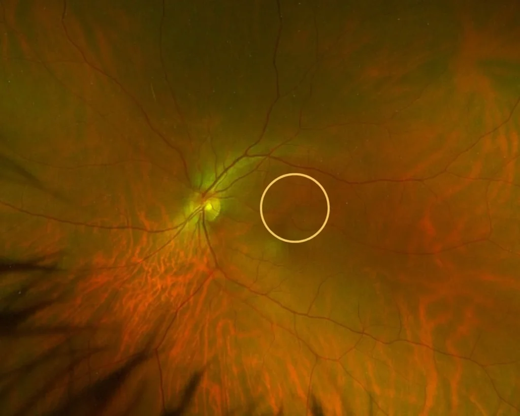
Your eyes can show more than how well you see. They can also show signs of disease. Many problems start before you feel pain. Often, your vision still seems normal. Because of this, imaging is very important. One tool doctors use is Optomap. It gives a wide, clear picture of the retina. The retina is the back part of your eye. This area connects to your brain. It also contains many small blood vessels.
With Optomap, doctors can see things you cannot feel or notice.
What Is Optomap?
Optomap is a type of retinal scan. It takes a large image of the back of your eye. In fact, it can capture up to 200 degrees in one shot. That is much wider than a regular exam.
Most eye tools show only a small section at a time. However, many diseases start in the outer edges. Those areas are hard to see without wide imaging. Because of this, small problems may be missed.
Optomap helps solve that problem. It gives a broad and detailed view.
How the Scan Works
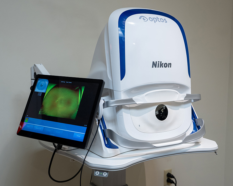
The test is simple. First, you sit in front of the machine. Then, you place your chin on a small rest. Next, you look at a light inside the device.
A quick flash scans your eye. The light is low powered and safe. The scan takes only seconds. After that, the image appears on a screen.
There is no pain. In most cases, you do not need dilation drops. As a result, you can return to normal activity right away.
Why You May Not Notice Eye Disease
Many eye diseases grow slowly. At first, they cause no pain. Vision often stays sharp. Because of this, people assume their eyes are healthy.
However, damage can begin quietly. For example, small blood vessels may leak. The optic nerve may slowly change. You cannot see or feel these shifts.
That is why imaging matters. It shows hidden changes early.
Finding Retinal Tears and Floaters
Floaters look like small dots or lines. They drift across your vision. Most floaters are harmless. Still, a sudden burst of floaters can mean trouble.
Sometimes, a retinal tear forms in the far edge of the eye. At first, you may not notice it. Yet the tear can grow fast.
Optomap captures the outer retina clearly. Because of this, doctors can spot small tears early. Early care helps prevent retinal detachment. That can save your vision.
Detecting Glaucoma Early
Glaucoma harms the optic nerve. It often develops slowly. Most people do not notice symptoms at first.
In many cases, side vision fades over time. However, central vision may stay clear. So the problem goes unnoticed.
Optomap shows the optic nerve in detail. Doctors look for subtle shape or color changes. If found early, treatment can begin sooner. Early treatment slows vision loss.
Monitoring Diabetic Retinopathy
Diabetes can damage tiny blood vessels in the retina. This condition is called diabetic retinopathy. Early stages often cause no symptoms.
Still, small leaks or swelling may begin. Without imaging, these changes are hard to see.
Optomap reveals these early signs. As a result, doctors can guide treatment sooner. Good control lowers the risk of vision loss.
Spotting Signs of Cancer
Some cancers can appear in the eye. Ocular melanoma is one example. It may start as a small dark spot.
Vision may seem normal at first. Even so, the growth can spread.
Optomap helps doctors detect unusual spots. If something looks abnormal, more testing can follow. Early action improves outcomes.
Seeing Signs of Stress and Blood Pressure
Stress affects the body in many ways. Over time, it can raise blood pressure. It can also strain blood vessels.
These changes sometimes appear in the retina. For instance, vessels may narrow or swell. Small bleeding spots may form.
Optomap shows these vessel patterns clearly. In some cases, an eye exam reveals health issues first.
High blood pressure also affects retinal vessels. Many people do not know they have it. Because symptoms are silent, damage can build slowly.
With retinal imaging, doctors may spot early warning signs. That can lead to faster medical care.
Why Optomap Is Helpful
Optomap does not replace a full exam. Instead, it adds more detail.
At first, it shows a wider area. Second, it stores digital images. Third, it allows year-to-year comparison.
This helps track small changes over time. Even tiny shifts become easier to notice.
Patients also benefit from seeing their own images. When people see the retina on screen, they understand their health better.
What Happens During Your Visit
The scan is quick. You sit comfortably. You look at a small light. Then the image is taken.
You may see a bright flash. It lasts less than a second. There is no contact with the eye.
After the scan, your doctor reviews the image. You will discuss any findings together.
Why Regular Scans Matter
Clear vision does not always mean healthy eyes. Many diseases stay silent for years.
Regular imaging helps doctors:
Watch the optic nerve
Check for retinal tears
Monitor diabetic changes
Track suspicious spots
Observe blood vessel patterns
Because images are saved, comparisons are easy. Early changes stand out more clearly.
A Simple Way to Protect Vision
Optomap is more than a camera. It is a tool for prevention. It helps doctors find problems early.
From glaucoma to retinal tears, many diseases begin without symptoms. The retina often shows the first clues.
When problems are found early, treatment works better. Vision loss can often be prevented.
Your eyes tell an important story. With wide imaging, doctors can read that story sooner. And that can protect both your sight and your health for years to come.
Schedule your appointment at 1 of our 4 conveniently located offices in San Jose, San Mateo, San Ramon, and Juneau.