
In the realm of eye care, early detection of eye diseases is paramount to preventing severe vision impairment or blindness. One groundbreaking technology that has revolutionized the field of optometry is Optomap retinal imaging. This non-invasive imaging technique allows eye doctors to capture a wide-field view of the retina, providing a comprehensive picture that aids in the early diagnosis and management of various eye conditions. The story behind the development of Optomap technology is as compelling as its contributions to eye health, rooted in a personal quest to prevent blindness, and is now credited with saving the lives of thousands of people every year.
The History of Optos
A Personal Quest
The inception of Optomap retinal imaging is a testament to the power of personal motivation and innovative thinking. The technology was developed by Douglas Anderson, a Scottish entrepreneur whose son, Leif, tragically went blind in one eye due to a retinal detachment that was not detected. At that time, it was difficult to examine retina health for young children and no technology was suited for use for children. Anderson’s determination to prevent other families from experiencing such a devastating loss drove him to create a better and safe method for examining the retina for young children.
Development and Evolution
In 1992, Anderson founded Optos plc with the vision of creating a device that could capture a detailed image of the retina without the need for dilation. His goal was to develop a technology that could provide a comprehensive view of the retina in a single capture, making it easier for eye care professionals to detect diseases early. After years of research and development, the first Optos device, the P200, was introduced in 2000. This pioneering device could capture a 200-degree view of the retina, a significant advancement compared to the traditional 45-degree view offered by conventional retinal cameras.
How Optomap Retinal Imaging Works
The Technology
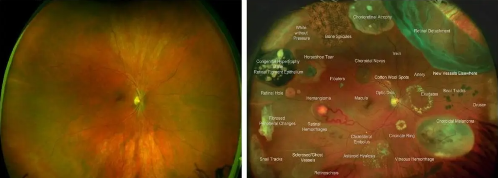
Optomap retinal imaging employs ultra-widefield (UWF) technology to capture high-resolution images of the retina. The device uses a low-power laser to scan the retina, creating a detailed image that includes the central and peripheral regions. This comprehensive view allows eye care professionals to examine the entire retina in a single image, providing critical information that can aid in the early detection of eye diseases.
The Process
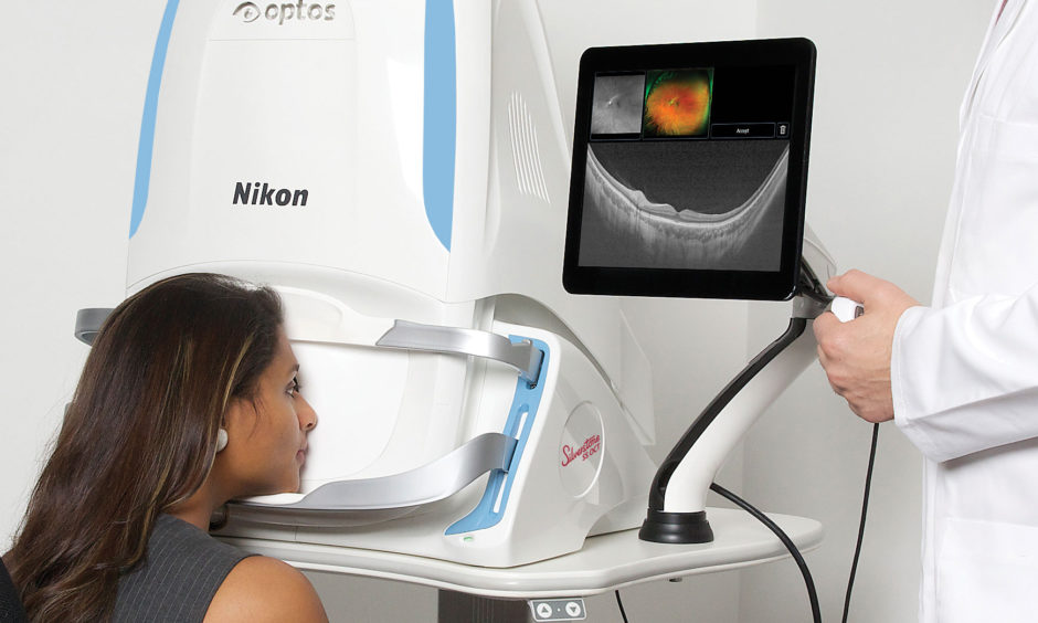
The imaging process is quick, non-invasive, and does not require dilation of the pupils, making it comfortable for patients. During the examination, the patient looks into the device, and within seconds, a high-resolution image of the retina is captured. This image can then be reviewed and analyzed by the eye doctor, providing valuable insights into the health of the retina.
Benefits of Optomap Retinal Imaging
Early Detection of Eye Diseases
One of the most significant benefits of Optomap retinal imaging is its ability to detect eye diseases at an early stage. Many eye conditions, such as diabetic retinopathy, glaucoma, and age-related macular degeneration (AMD), develop gradually and may not present noticeable symptoms until they are advanced. By capturing a detailed image of the retina, Optomap technology allows eye care professionals to identify early signs of these diseases and initiate timely treatment.
- Diabetic Retinopathy – Diabetic retinopathy is a leading cause of blindness in adults. It occurs when high blood sugar levels cause damage to the blood vessels in the retina. Optomap retinal imaging can detect early signs of diabetic retinopathy, such as microaneurysms, hemorrhages, and exudates, allowing for early intervention and better management of the condition.
- Glaucoma – Glaucoma is a group of eye conditions that damage the optic nerve, often due to increased intraocular pressure. Early detection is crucial in preventing vision loss from glaucoma. Optomap imaging can reveal changes in the optic nerve head and retinal nerve fiber layer, which are indicators of glaucoma, enabling prompt treatment to preserve vision.
- Age-Related Macular Degeneration (AMD) – AMD is a common condition that affects the macula, the part of the retina responsible for central vision. Early detection of AMD is vital for preserving vision and slowing the progression of the disease. Optomap retinal imaging can identify early signs of AMD, such as drusen (yellow deposits) and changes in the retinal pigment epithelium, facilitating early management and treatment.
- Retinal Tears and Retinal Detachments – Widefield optomap retinal imaging is one of the best ways to supplement a dilation when patients experience flashes and floaters, one of the top symptoms for a retinal tear and detachment. Being able to see up to 92% of the retina all at once allows eye doctors to diagnose and refer quickly to a retinal specialist if an issue is found.
Comprehensive Examination with Optomap retinal imaging
The ultra-widefield view provided by Optomap retinal imaging offers a more comprehensive examination of the retina compared to traditional methods. By capturing a 200-degree view, eye care professionals can assess the peripheral regions of the retina, which are often missed during routine examinations. This comprehensive view is particularly beneficial for detecting peripheral retinal tears, detachments, and lesions that may not be visible with conventional imaging techniques.
Improved Patient Experience
Optomap retinal imaging enhances the patient experience by providing a quick and comfortable examination process. Since dilation is not required, patients can avoid the discomfort and inconvenience associated with dilated eye exams. The non-invasive nature of the procedure also makes it suitable for patients of all ages, including children and individuals with light sensitivity.
Enhanced Monitoring and Documentation
Optomap retinal imaging allows for detailed documentation of the retina, which is essential for monitoring changes over time. By comparing images from different visits, eye care professionals can track the progression of eye conditions and assess the effectiveness of treatment. This capability is particularly valuable for managing chronic eye diseases and making informed decisions about patient care.
The Role of Optomap in Modern Optometry
Integrating Optomap Technology in Clinical Practice
The adoption of Optos retinal imaging has become increasingly prevalent in modern optometry practices. Eye care professionals recognize the value of this technology in enhancing diagnostic capabilities and improving patient outcomes. By incorporating Optos imaging into routine eye exams, optometrists can provide a higher standard of care and offer their patients the best possible protection against vision loss.
Advancements in Optomap Retinal Imaging
Since the introduction of the first Optos device, the technology has continued to evolve. Newer models, such as the Monaco and Silverstone, offer even higher resolution imaging and additional features, such as autofluorescence and angiography, to provide more comprehensive retinal assessments. These advancements further enhance the ability of eye care professionals to detect and manage a wide range of eye conditions.
Research and Education
Optomap retinal imaging has also contributed significantly to research and education in the field of optometry. The detailed images captured by Optos devices provide valuable data for clinical studies, helping researchers better understand the progression of eye diseases and develop new treatment strategies. Additionally, the technology serves as an educational tool for optometry students and practitioners, allowing them to study the retina in greater detail and improve their diagnostic skills.
Schedule an Exam with Optomap Retinal Imaging
Optomap retinal imaging represents a major advancement in the field of optometry, offering a powerful tool for the early detection and management of eye diseases. The technology’s ability to capture a comprehensive view of the retina, coupled with its non-invasive and patient-friendly nature, makes it an invaluable asset in modern eye care. The compelling story behind its development, driven by Douglas Anderson’s determination to prevent blindness, underscores the importance of innovation in improving patient outcomes. By embracing Optos technology, eye care professionals can provide their patients with the highest standard of care, helping to preserve vision and prevent blindness.
At Optical Illusions: An Optometric Practice, we are committed to providing exceptional eye care. Our experienced optometrists use state-of-the-art technology to conduct thorough retinal screenings and comprehensive eye exams. Don’t wait until symptoms appear—schedule your eye exam today and take proactive steps towards safeguarding your vision and health. Contact our team to schedule your appointment at 1 of our 4 conveniently located offices.